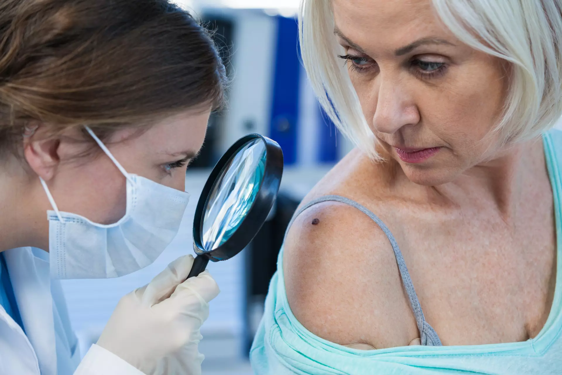
Most moles are completely benign — clusters of pigment-producing cells that develop during childhood and early adulthood and remain stable for decades. The vast majority will never cause any problem. But melanoma, the most serious form of skin cancer, often begins in or near a mole. The ability to distinguish between the two is not something that can be reliably done in a bathroom mirror.
At South Florida Skin Center, mole evaluations are done with dermoscopy — a magnified imaging technique that reveals the internal structure of pigmented lesions in a way that significantly extends what clinical examination alone can assess. Features like the regularity of pigment networks, the presence of atypical vascular patterns, and the characteristics of the lesion’s border all inform whether something warrants monitoring, biopsy, or removal. If a lesion looks atypical, we tell you what we’re seeing, why it concerns us, and what a biopsy would tell us. We don’t hedge.
The ABCDE rule — a useful starting point
The ABCDE criteria give patients a framework for self-monitoring between professional exams: Asymmetry (one half doesn’t match the other), Border irregularity (ragged, notched, or blurred edges), Color variation (multiple shades of brown, black, red, white, or blue within a single lesion), Diameter over 6mm (roughly the size of a pencil eraser), and Evolution (any change in size, shape, color, or new symptoms like itching or bleeding).
These criteria are helpful but imperfect. Some early melanomas are small, symmetric, and uniform in color. Others are amelanotic — lacking the dark pigmentation patients typically associate with a dangerous mole — and can be missed without dermoscopic evaluation. Professional assessment adds something self-examination cannot.
When removal makes sense
Not every mole needs to come off. Many atypical-appearing moles can be safely monitored with serial dermoscopic photography, which documents the baseline appearance and allows us to detect change over time before it reaches a clinically dangerous threshold.
When removal is appropriate — because a lesion looks suspicious, has changed, is symptomatic, or the patient simply prefers not to monitor it indefinitely — the method depends on what the lesion is and where it sits. Shave removal is appropriate for raised, clearly benign lesions. Surgical excision is used for lesions that require complete removal and margin assessment. We don’t apply one approach to every mole.
For lesions removed with concern about malignancy, the specimen goes to dermatopathology. We review the results with you and discuss what they mean for follow-up.
Frequently asked questions
Does mole removal hurt?
The procedure is done under local anesthesia. Most patients feel pressure more than pain. There’s typically some soreness at the site for a day or two afterward. The anticipation is usually worse than the experience.
Will it leave a scar?
Any time the skin is broken, some degree of marking is possible. What the scar looks like depends on the method, the location on the body, and how your skin heals. We discuss this honestly before any procedure. Shave removals typically heal with minimal scarring. Excisional removal leaves a linear scar whose final appearance improves significantly over 12 to 18 months.
I have a lot of moles. Should I have them all evaluated?
A full-body skin exam is the practical way to evaluate all of them at once. We document any moles that warrant monitoring and establish a baseline for comparison at future visits. For patients with many atypical-appearing moles, we often use dermoscopic photography to create a formal record.
When is the right time to come in?
If a mole is changing — growing, darkening, developing irregular borders, bleeding, or simply looking different from before — don’t wait for your next annual exam. That is specifically the scenario that benefits from prompt evaluation.